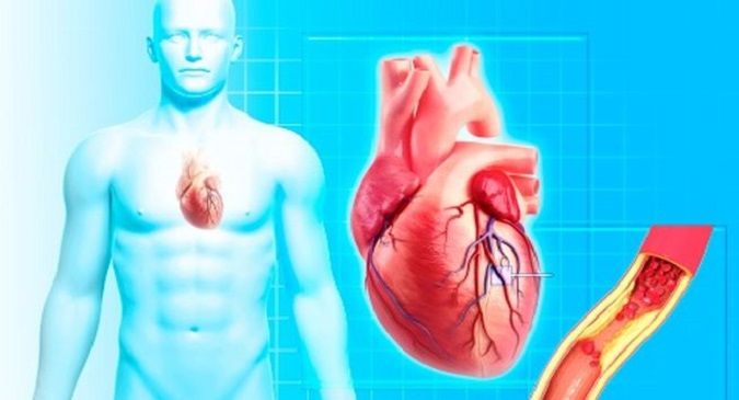
Rheumatic heart disease (RHD) remains a leading cause of cardiovascular morbidity in children and young adults in many low- and middle-income regions. Despite being largely preventable, RHD continues to exact a heavy toll on public health. At First Point MD, we believe that understanding the interplay between valve involvement, accurate diagnosis, proactive management, and prevention strategies is essential if we are to reduce the global burden of this disease. In this comprehensive article, we explore the mechanisms, diagnosis, valvular pathology, therapeutic interventions, and prevention measures vital to controlling RHD.
Understanding Rheumatic Heart Disease: Pathogenesis and Epidemiology
RHD is not an infection of the heart per se, but rather a post-infectious immunologic sequela following group A Streptococcus (GAS) infection—typically of the throat (streptococcal pharyngitis). In susceptible individuals, immune cross-reactivity leads to inflammation in the heart, joints, skin, and central nervous system, manifesting as acute rheumatic fever (ARF). Repeated or severe episodes of ARF injure the cardiac valves, leading over time to fibrosis, scarring, and deformity—the hallmarks of RHD.
Globally, RHD affects over 50 million people and causes approximately 360,000 deaths annually, especially in low-resource settings. The prevalence is highest in parts of Sub-Saharan Africa, South Asia, the Pacific, and marginalized communities. Socioeconomic determinants—overcrowding, poor sanitation, limited healthcare access—play a central role in sustaining GAS transmission and undertreatment of pharyngitis.
Valve involvement is the core of RHD’s clinical significance. Typically, left-sided valves are predominantly affected (mitral > aortic). Less often, the tricuspid or pulmonary valves may be involved, usually in association with mitral disease.
Valve Pathology in RHD: What Happens to the Heart Valves
The hallmark of RHD is structural damage to cardiac valves. Key pathological features include:
-
Inflammation and valvulitis during the acute ARF phase
-
Fibrosis, thickening, calcification, and leaflet fusion in chronic phases
-
Chordae tendineae fusion or shortening
-
Commissural fusion (especially in mitral stenosis)
-
Restricted leaflet mobility and regurgitant jets
Mitral regurgitation (MR) is the most common early lesion; over time, mitral stenosis (MS) may evolve, often via progressive thickening and commissural fusion. Aortic regurgitation or stenosis may co-exist, though less frequently. In severe disease, mixed valvular lesions often complicate surgical planning.
Valvular damage reduces valve function, leading to volume overload, pressure overload, chamber remodeling, atrial enlargement, and eventually heart failure, arrhythmias (particularly atrial fibrillation), and pulmonary hypertension. Recognizing early valve involvement before irreversible changes is critical to preserving long-term cardiac function.
Clinical Presentation: Recognizing RHD and Valve Dysfunction
RHD often evolves silently, with symptoms appearing only after significant valve damage. Common clinical features include:
-
New or changing heart murmurs (especially pansystolic, diastolic, or mid-diastolic murmurs)
-
Dyspnea, particularly on exertion or when progressing to heart failure
-
Fatigue, weakness, reduced exercise tolerance
-
Palpitations, syncope (often secondary to atrial arrhythmias)
-
Edema, ascites, hepatic congestion when heart failure is advanced
In children or young adults with acute rheumatic fever, systemic features like arthritis, migratory joint pain, fever, chorea, erythema marginatum, or subcutaneous nodules may precede heart symptoms.
A key clinical red flag is a new murmur or change in character of an existing murmur in someone with a history of streptococcal infection or rheumatic fever. Close attention to such changes can prompt timely investigation.
Diagnosis: Evaluating Valve Involvement and Disease Severity
Comprehensive and precise diagnosis of RHD hinges on a combination of clinical evaluation, laboratory testing, and imaging modalities.
Clinical and Laboratory Evaluation
-
Detailed history and physical examination: Assessing for signs of valvular dysfunction, prior streptococcal infections, or ARF episodes
-
Electrocardiogram (ECG): To detect arrhythmias (e.g. atrial fibrillation), conduction abnormalities, atrial enlargement
-
Chest radiography (X-ray): To assess cardiomegaly, pulmonary vascular congestion
-
Inflammatory markers (e.g., ESR, CRP) and antistreptococcal serologies (e.g. anti-streptolysin O titer) during acute phases
However, neither ECG nor X-ray can reliably detect early valvular involvement or the subtle structural changes of RHD.
Echocardiography: The Diagnostic Gold Standard
Transthoracic echocardiography (TTE) is the backbone of RHD diagnosis. It detects morphological and functional derangements in valve structure and flow dynamics with high sensitivity and specificity.
The World Heart Federation (WHF) criteria for echocardiographic diagnosis of RHD (especially in children ≤20 years) provide standardized definitions of definite, borderline, or normal disease, based on morphologic features (leaflet thickening, restricted motion, chordal thickening) and pathologic regurgitation or stenosis parameters.
Findings such as “hockey-stick” mitral leaflet motion, commissural fusion, or thickened chordae suggest rheumatic etiology.Subclinical RHD may be detected by echo even in absence of auscultatory murmurs—underscoring the power of screening echocardiography in endemic settings.
In some advanced cases, transesophageal echocardiography (TEE) or 3D echo may further clarify valve anatomy, especially prior to surgical or percutaneous intervention.
Other Imaging and Novel Diagnostics
-
Cardiac MRI might help assess chamber remodeling and myocardial fibrosis, though its role in RHD is limited due to cost and accessibility
-
Machine learning approaches using ECG and phonocardiographic (PCG) signal analysis have emerged in research settings for RHD screening, particularly in low-resource settings aiming to bypass the lack of echocardiography.
-
Doppler studies quantify regurgitant volumes, gradients, and valve areas, essential for staging severity
Grading Valve Dysfunction
Once valve involvement is confirmed, severity must be graded (e.g. mild, moderate, severe) based on standard measures—regurgitant volume, effective regurgitant orifice area, pressure gradients, valve area in stenosis, and hemodynamic consequences. This informs decision-making about interventions.
Management: Medical, Interventional, and Surgical Strategies
Effective care of RHD demands a multidisciplinary approach combining medical therapy, secondary prophylaxis, valve interventions, and long-term monitoring.
Medical Therapy and Symptom Relief
-
Heart failure management (where present): use of ACE inhibitors, ARBs, diuretics, beta-blockers, and afterload reduction (depending on valvular lesion)
-
Rate control or anticoagulation for atrial fibrillation
-
Endocarditis prophylaxis: while earlier guidelines recommended long-term prophylactic antibiotics for RHD patients during dental procedures, contemporary guidelines have narrowed indications per newer AHA recommendations; RHD alone is no longer automatically an indication for prophylaxis.
However, these medical measures do not reverse valve disease—they are palliative and supportive.
Secondary Prophylaxis: Preventing Recurrence of ARF
Crucially, continuous antibiotic prophylaxis is the keystone of preventing further valvular damage. Benzathine penicillin G, given intramuscularly every 3–4 weeks, is the standard. In some high-risk settings, every 3 weeks is favored.
Duration varies—from 5–10 years or until age 21–40, and for severe RHD or after valve surgery, lifelong prophylaxis may be warranted. The precise optimal duration remains debated.
Secondary prophylaxis is supported by both WHO recommendations and national guidelines as the most cost-effective strategy to halt disease progression.
Percutaneous and Surgical Valve Interventions
Once valve disease is advanced or symptomatic beyond medical therapy, structural interventions are crucial:
-
Percutaneous mitral balloon valvuloplasty (PMBV / percutaneous mitral balloon commissurotomy) is first-line for many cases of mitral stenosis without heavy calcification, significant regurgitation, atrial thrombus, or other contraindications.
-
Valve repair or valve replacement (mechanical or bioprosthetic) become necessary in severe lesions of the mitral and/or aortic valves.
-
Surgical timing is critical: delaying until irreversible ventricular dysfunction or pulmonary hypertension sets in increases risk.
-
Even after surgical correction, secondary prophylaxis must continue to prevent reinjury of prosthetic or repaired valves.
Choice of intervention depends on patient factors (age, comorbidities), valve morphology, resource availability, and surgical expertise.
Prevention: The Best Strategy Is to Prevent Disease
Because damage from RHD is irreversible, prevention is the most powerful intervention. Prevention is three-tiered:
Primary Prevention (Prevent ARF)
-
Early diagnosis and antibiotic treatment of GAS pharyngitis (e.g. penicillin or amoxicillin) to prevent initial ARF.
-
In high-burden settings, empirical treatment of sore throats when clinical likelihood is high may be justified in absence of testing.
-
Population-level strategies: health education, improved hygiene, reducing overcrowding, enhancing access to medical care.
Secondary Prevention (Prevent Recurrence)
-
As discussed above, continuous prophylactic antibiotics (IM benzathine penicillin G) to interrupt repeat GAS infections and recurrent ARF, thereby preventing further valvular damage.
-
Ensuring a consistent supply chain for penicillin is key, as global shortages have disrupted care in endemic regions.
-
Community-based registry and recall systems help monitor compliance and detect defaults early.
Tertiary Prevention (Manage Established RHD)
-
Early detection of valve lesions through echocardiographic screening and close follow-up
-
Timely surgical or percutaneous intervention before irreversible damage
-
Optimal medical management of complications (heart failure, arrhythmias)
-
Lifelong follow-up and secondary prophylaxis
Challenges, Gaps, and the Way Forward
While the framework of diagnosis, management, and prevention is well known, many real-world challenges impede progress:
-
Underdiagnosis and Late Presentation: Many patients present only when symptoms are advanced, limiting salvageability.
-
Limited Access to Echocardiography and Specialist Care in low-resource settings
-
Poor Adherence to Prophylaxis, owing to cost, access, fear of injections, or unawareness
-
Antibiotic Supply Disruptions—especially for benzathine penicillin
-
Uncertain Duration of Prophylaxis, particularly in varying risk strata
-
Need for Innovation: point-of-care diagnostics, machine learning–aided screening, vaccine development against GAS are under exploration.
To overcome these gaps, integrated programs combining community engagement, public health infrastructure, surveillance registries, and sustainable funding are essential.
Closing Thoughts
Rheumatic heart disease remains a preventable tragedy. While valve involvement is the central pathology driving morbidity and mortality, our success lies in recognizing disease early, managing it judiciously, and preventing its onset altogether. At First Point MD, we are committed to:
-
Promoting standardized echocardiographic screening and diagnostic vigilance
-
Implementing rigorous secondary prophylaxis regimens
-
Ensuring access to valve interventions before irreversible damage
-
Collaborating with public health agencies to bolster primary prevention
With concerted global effort, we can reduce the toll of RHD and give countless individuals a chance at healthier hearts and better futures.


